This is the list of FISH tests available in the laboratory.
Click on the gene name for general information of the probe and its respective common applications. Web-links to the manufacturers' websites and genomic maps of each of the loci are provided.
Please note that only ALK breakapart FISH and ROS1 breakapart FISH are accrediated tests.
Please order the test through the desktop little fox.
FISH is performed on formalin-fixed paraffin embedded tissue sections. Composed-pseudocoloured images are captured for enumeration. Pathologists might count at least 50 interphase tumor nuclei. Positive cut-off for FISH tests varies on the nature of probes, specific genomic changes and in different diseases. As low as 15% has been proposed in published guidelines for some probes e.g. some types of BRAF translocation in pilocytic astrocytoma, RET translocations in non small cell lung carcinomas, etc.
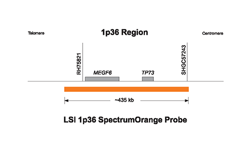
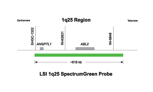
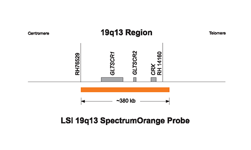
Vysis LSI 1p36 SpectrumOrange/1q25 SpectrumGreen Probes and Vysis LSI 19q13 SpectrumOrange/19p13 SpectrumGreen Probes (ASR) is used in 2 separated FISH tests. Deletions affecting the short arm of chromosome 1 (1p36) and the long arm of chromosome 19 (19q13) are frequently found in human gliomas and are associated with a better survival. Determination of 1p and 19q status may aid in therapeutic decisions and predict outcome in patients with anaplastic oligodendrogliomas.
Please use Ronnie's template for scoring of a total 200 tumor nuclei.
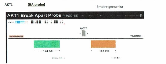
Orange Probe: centromere side, AKT1 3' end
Green Probe: telomere side, AKT1 5'end
Empire Genomics’ AKT1 Break Apart FISH Probe is designed to flank the AKT1 gene and is typically used for detecting AKT1 rearrangements such as translocations.
For Non-small cell lung carcinoma (CAP accredited test).
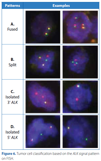
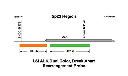
The Vysis ALK Break Apart FISH Probe Kit is a qualitative test to detect rearrangements involving the ALK gene via fluorescence in situ hybridization (FISH) in formalin-fixed paraffin-embedded (FFPE) non-small cell lung cancer (NSCLC) patients. See also "IASLC Atlas of ALK adn ROS1 testing in lung cancer 2e". Special note is to be taken if there are solely 5' signals (isolated green signals).
Orange Probe: Telomere side, 3' end of the ALK gene.
Green Probe: 3' end to mid portion of the ALK gene (including the tyrosine kinase domain).
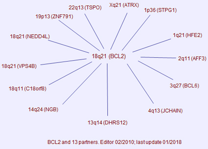
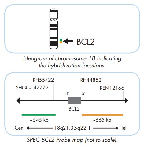
For B-cell lymphoma, follicular lymphoma.
Orange Probe: telomere side, BCL2 5' end
Green Probe: centromere side, BCL2 3' end
The ZytoLight ® SPEC BCL2 Dual Color Break Apart Probe is designed to detect translocations involving the chromosomal region 18q21.33 harboring the BCL2 gene. Translocations involving the BCL2 gene are commonly identified in B-cell lymphomas. In particular, the translocation t(14;18)(q32.3;q21.3) has been identified in about 80% of follicular lymphoma.
adapted from "Atlas of Genetics and Cytogenetics in Oncology and Haematology"
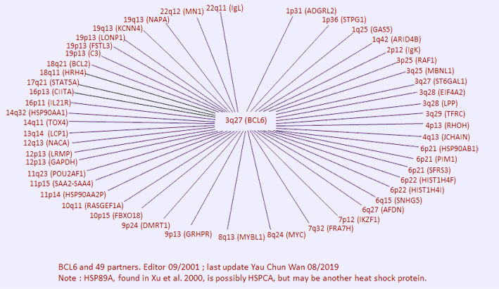
For double hit, triple hit diffuse large B-cell lymphoma.
Orange Probe: telomere side, BCL6 5' end
Green Probe: centromere side, BCL6 3' end
Vysis LSI BCL6 (ABR) Dual Color Break Apart DNA probe hybridizes to the band 3q27. The 5’ BCL6 SpectrumOrange probe is ~349 kb in size and flanks the ABR of BCL6. The 3’ BCL6 SpectrumGreen probe is approximately 816kb in size and flanks the MBR region of BCL6. There is an approximate 265 kb gap between the two probes. In interphase nuclei of normal cells, the probe generally appears as two distinct signals (orange and green adjacent, or fused yellow). Probe signals may also appear diffuse or split in interphase nuclei, depending upon the condensation of the DNA.
In high-grade B-cell lymphomas, the presence of MYC aberrations identifies a patient subset with a very poor prognosis, particularly when there is concomitant rearrangement of BCL2 or BCL6, a condition referred to as "double hit” DLBCL. In rare cases translocation involves MYC, BCL2 and BCL6, so called “triple hit”.
adapted from "Atlas of Genetics and Cytogenetics in Oncology and Haematology"
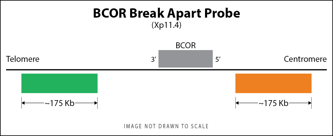
for Ewing sarcoma-like small blue round cell tumors, endometrial stromal sarcoma.
Red Probe : centromere side, 3' of the BCOR gene
Green Probe : telomere side, 5' of the BCOR gene
Fluorescence in situ hybridization (FISH) using GSP BCOR Probe (Guangzhou Lbp Medicine Science & Technology Co., Ltd.) and is typically used for detecting BCOR rearrangements such as translocations in small blue round cell tumors, Ewing sarcoma-like tumors that are negative for EWSR1 translocations.
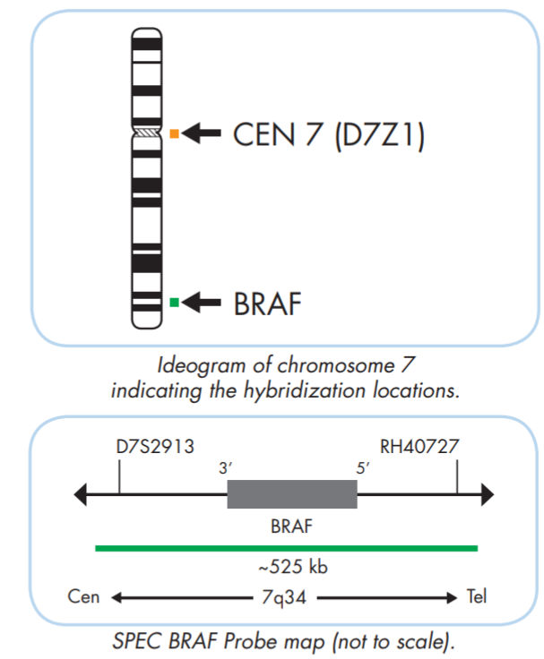
for brain tumomr typing.
Orange Probe: CEN7 (D7Z1)
Green Probe: BRAF, 7q34, ~525 kb
The ZytoLight ® SPEC BRAF/CEN 7 Dual Color Probe is designed for the detection of amplifications involving the chromosomal region 7q34 harboring the BRAF gene. Activating mutations in BRAF are found in many tumor types, including malignant melanoma, thyroid, colorectal, and ovarian carcinomas, lung adenocarcinoma, as well as in some sarcomas and gliomas.
BRAF/CEN7 amplification probe might also be used to look for KIAA-BRAF translocation in pilocystic astrocytoma, consisting of complex duplication and translocation events. RT-PCR assay might, sometimes, be more efficient in detecting this particular translocation.
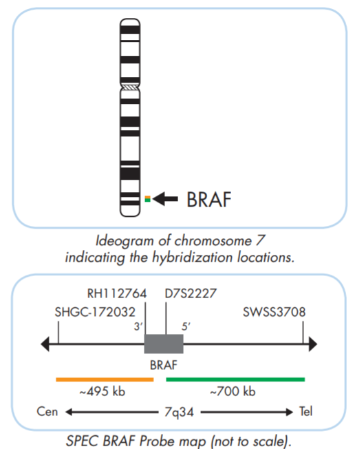
for brain tumomr typing.
Orange Probe: centromere side, BRAF 3' end
Green Probe: telomere side, BRAF 5'end
The ZytoLight ® SPEC BRAF Dual Color Break Apart Probe is designed for the detection of rearrangements involving the chromosomal region 7q34 harboring the BRAF gene. Various BRAF translocations were observed in melanocytic nevi, pilocytic astrocytomas, malignant melanoma, prostate and gastric cancer.
adapted from "Atlas of Genetics and Cytogenetics in Oncology and Haematology"
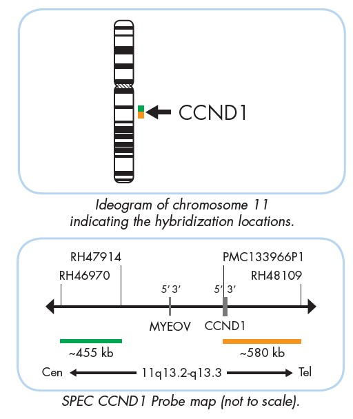
For mantle cell lymphoma
Orange Probe: telomere side, CCND1 3' end
Green Probe: centromere side, CCND1 5' end
The ZytoLight SPEC CCND1 Dual Color Break Apart Probe (PL65) is intended to be used for the qualitative detection of translocations involving the human CCND1 gene at 11q13.3 in formalin-fixed, paraffin-embedded specimens by fluorescence in situ hybridization (FISH).
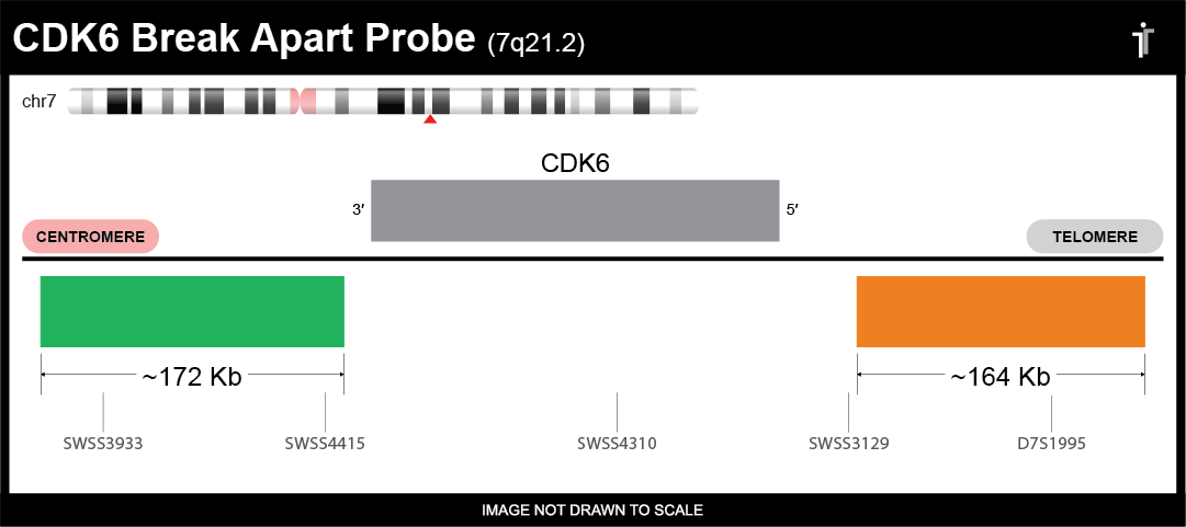
Orange Probe: telomere side, CDK6 5' end
Green Probe: centromere side, CDK6 3' end
Empire Genomics’ CDK6 Break Apart FISH Probe is designed to flank the CDK6 gene and is typically used for detecting CDK6 rearrangements such as translocations.
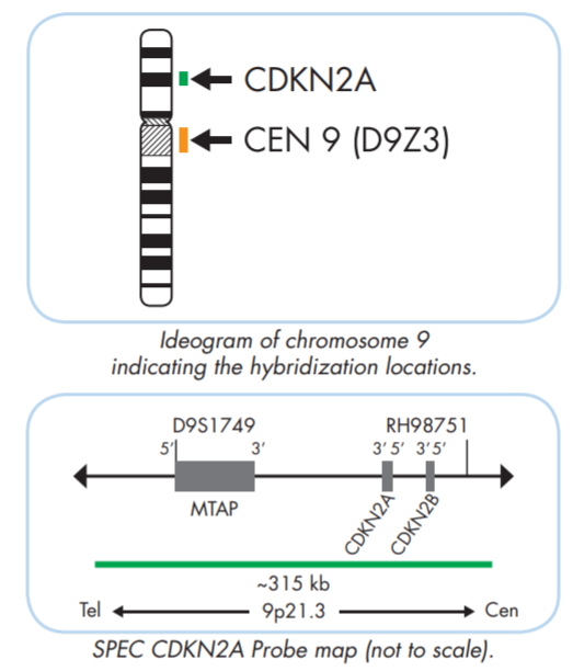
for brain tumor typing.
Orange Probe: CEN 9 (D9Z3)
Green Probe: CDNK2A, 9p21.3, ~315 kb
The ZytoLight ® SPEC CDKN2A/CEN 9 Dual Color Probe is designed for the detection of CDKN2A deletions frequently observed in most tumor cell lines as well as in primary human malignancies. CDKN2A gene is found in up to 80% of T-cell acute lymphoblastic leukemia cases and is associated with poor prognosis and relapse of the disease. Also, this genetic change might be important in the classification and acts as prognostic indicator for some gliomas, aiming to look at homozygous deletion.
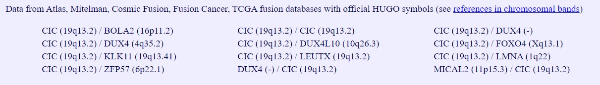
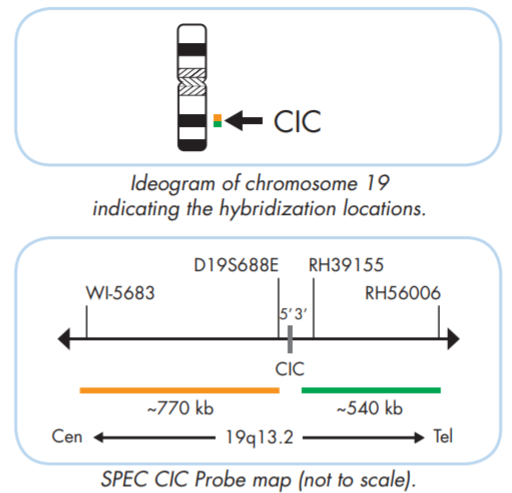
for Ewing sarcoma-like small blue round cell tumors.
Orange Probe: centromere side, CIC 5' end
Green Probe: telomere side, CIC 3' end
The ZytoLight ® SPEC CIC Dual Color Break Apart Probe is designed to detect translocations involving the chromosomal region 19q13.2 harboring the CIC gene. Rearrangements involving the CIC gene are frequently found in EWSR1-negative small blue round cell tumors and have been described as aggressive tumors with an inferior overall survival compared to Ewing sarcoma.
adapted from "Atlas of Genetics and Cytogenetics in Oncology and Haematology"
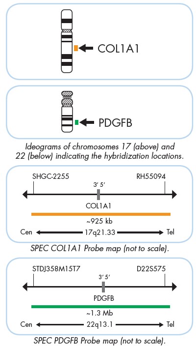
for dermatofibrosarcoma protuberans (DFSP).
Orange Probe: COL1A1, 17q21.33, ~925 kb
Green Probe: PDGFB, 22q13.1, ~1.3 Mb
The ZytoLight ® SPEC COL1A1/PDGFB Dual Color Dual Fusion Probe is designed for the detection of the specific translocations involving the chromosomal region 17q21.33 harboring the COL1A1 (a.k.a. OI4) gene, and the chromosomal region 22q13.1, harboring the PDGFB (a.k.a PDGF2, SIS) gene. The reciprocal translocations involving t(17;22)(q21.3;q13.1) are characteristic for dermatofibrosarcoma protuberans (DFSP) patients.
Local lab data criteria: POSITIVE (>=20%), NEGATIVE (<10%), and further RT-PCR for cases with 10-20% positive nuclei. (This is our first fusion probe, please comment.)
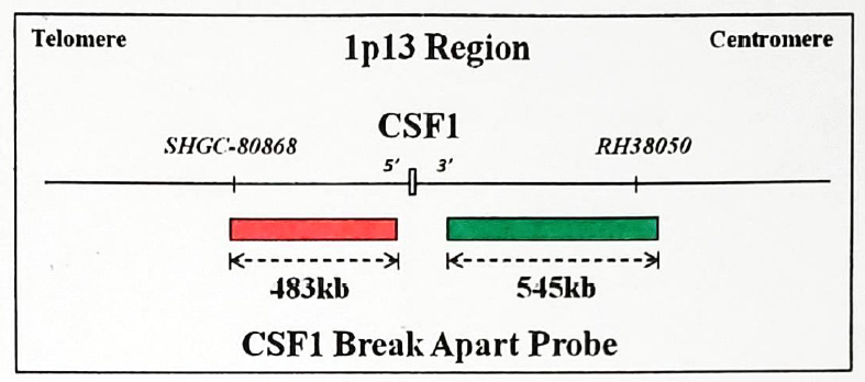
for PVNS and TGCT. Probe from "Guangzhou Lbp Medicine Science & Technology Co., Ltd."
Orange Probe: telomere side, CSF1 5' end
Green Probe: centromere side, CSF1 3' end
CSF1, the ligand of the tyrosine kinase receptor, CSF1R, can be translocated in pigmented villonodular synovitis (PVNS) and tenosynovial giant cell tumor (TGCT). Am J Surg Pathol. 2007 Jun;31(6):970-6.
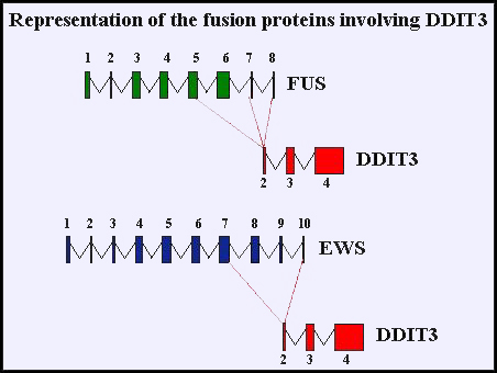
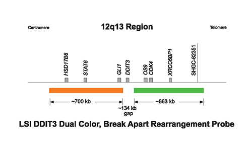
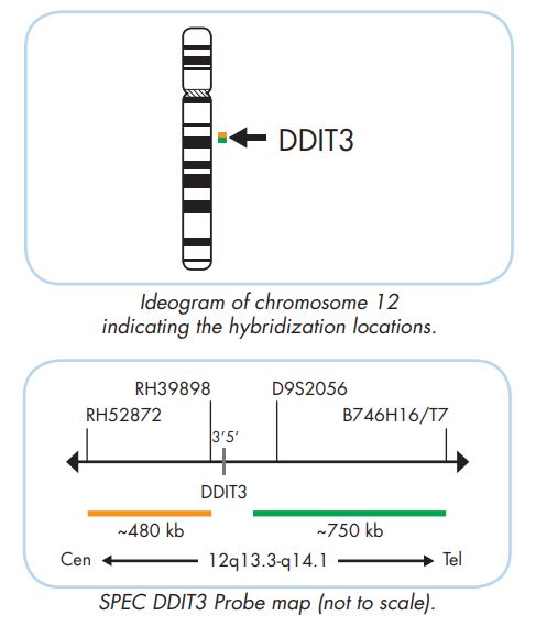
for myxoid liposarcoma.
Orange Probe: centromere side, DDIT3 3' end
Green Probe: telomere side, DDIT3 5' end
Vysis DDIT3 Break Apart FISH Probe Kit and the ZytoLight ® SPEC DDIT3 Dual Color Break Apart Probe are designed to detect translocations involving the chromosomal region 12q13.3 harboring the DDIT3 gene. DDIT3 is consistently rearranged in myxoid liposarcomas.
adapted from "Atlas of Genetics and Cytogenetics in Oncology and Haematology"
Vysis Probe
ZytoLight Probe
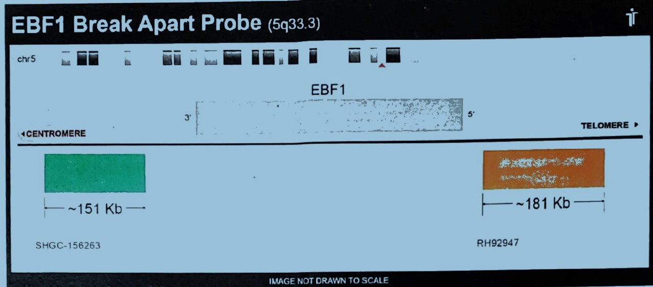
Orange Probe: telomere side, EBF1 5' end
Green Probe: centromere side, EBF1 3' end
For sarcoma, such as lipomatous neoplasm and lipofibrous neoplasm. Pediatric B-cell precursor acute lymphoblastic leukemia (BCP-ALL)
adapted from "Atlas of Genetics and Cytogenetics in Oncology and Haematology"
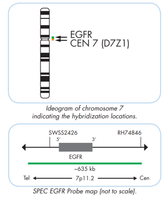
for brain tumomr typing.
Orange Probe: CEN 7 (D7Z1)
Green Probe: EGFR, 7p11.2, ~635 kb
The ZytoLight ® SPEC EGFR/CEN 7 Dual Color Probe is designed for the detection of EGFR gene amplification frequently observed in solid neoplasms including non-small-cell lung cancer (NSCLC) and glioblastoma. Overexpression of EGFR has been shown in a number of tumor entities and is associated with poor prognosis. This probe is also used in the diagnostic algorithm of glioma.
A tumor was considered EGFR amplified when there was focal EGFR gene amplification defined as an EGFR/CEP 7 ratio greater than or equal to 2 in ≥15% recorded cells. Tumors with polysomy for chromosome 7 (excess copies of the entire chromosome) but without focal amplification of the EGFR gene were considered to be EGFR nonamplified. (French et al 2020 Neuro-Oncol)
Please order HER2 DISH for routine breast CA biomarker testing.
The PathVysion HER-2 DNA Probe Kit (PathVysion Kit) is designed to detect amplification of the HER-2/neu gene via fluorescence in situ hybridization (FISH) in formalin-fixed, paraffin-embedded human breast cancer tissue specimens. We have taken off this test from accreditation. Please order DISH through the desktop little fox for more efficient processing in immuno lab using the automated processor.
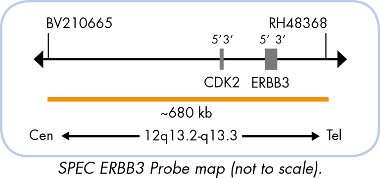
Orange Probe: ERBB3, 12q13.2-q13.3, ~680 kb
Green Probe: CEN12 (D12Z3)
The ZytoLight ® SPEC ERBB3/CEN 12 Dual Color Probe is designed for the detection of amplifications of the chromosomal regions harboring the ERBB3 gene. It belongs to the epidermal growth factor receptor subgroup of the receptor tyrosine kinase superfamily which is known to be affected by gene amplifications in a number of malignant tumors.
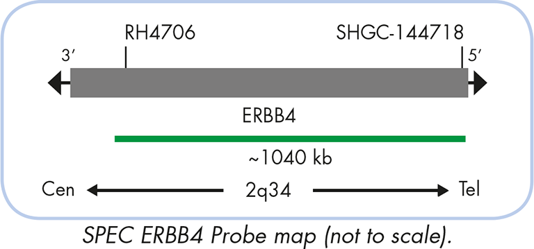
The ZytoLight ® SPEC ERBB4/2q11 Dual Color Break Apart Probe is designed for the detection of amplifications of the chromosomal regions harboring the ERBB4 gene. There is growing evidence that cooperation of all four members of the ERBB gene family contributes to a more aggressive tumor phenotype and influences therapeutic response.
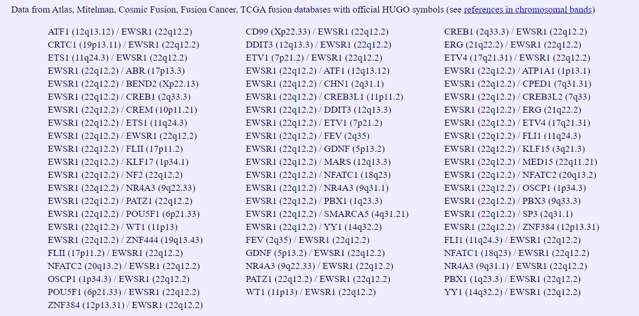
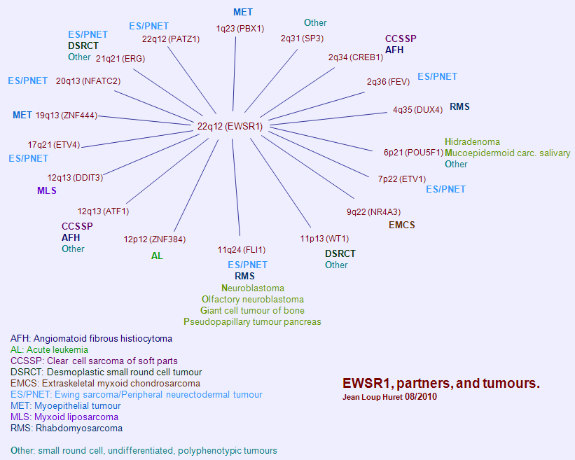
for many different sarcomas including angiomatoid fibrous histiocytoma, Clear cell sarcoma of soft parts, desmoplastic small round cell tumor, extraskeletal myoxoid chondrosarcoma, Ewing sarcoma/PNET, myoepithelial tumor, .....
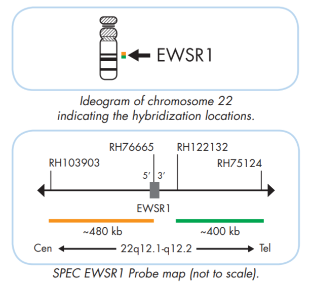
Orange Probe: centromere side, EWSR1 5' end
Green Probe: telomere side, EWSR1 3' end
The ZytoLight ® SPEC EWSR1 Dual Color Break Apart Probe is designed to detect translocations involving the chromosomal region 22q12.2 harboring the EWSR1 gene (Ewing sarcoma breakpoint region 1). There are many fusion partners for the EWSR1 translocation.
Sanger's Catalogue of Somatic Mutations In Cancer (COSMIC) fusion database provides an updated references of the breakpoints for EWSR1.
adapted from "Atlas of Genetics and Cytogenetics in Oncology and Haematology"
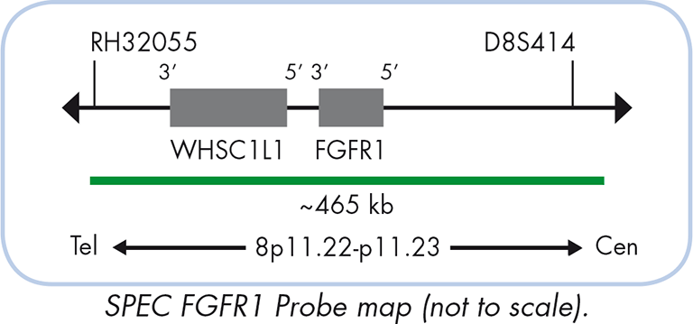
The ZytoLight ® SPEC FGFR1/CEN 8 Dual Color Probe is designed for the detection of FGFR1 gene amplification frequently observed in malignant tumors e.g. breast and prostate cancer and oral squamous cell carcinoma (OSCC). FGFR1 is believed to emerge as a potential therapeutic target for lobular breast carcinomas.
for calcifying aponeurotic fibroma (FN1 and EGF fusion)
for chondroblastoma (FN1 and FGFR1/FGFR2 fusion)
for chondroid and calcified mesenchymal neoplasms (FN1 and FGFR2, FGFR1, MERTK, NTRK1, TEK fusions)
for phosphaturic mesenchymal tumor (FN1 and FGFR1/FGF1 fusion)
for soft tissue chondromas (FN1 and FGFR2 fusion)
for synovial chondromatosis (FN1 and ACVR2 fusions).
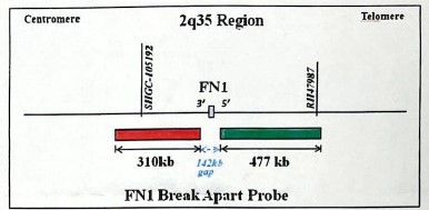
Orange Probe: telomere side, FN1 5' end
Green Probe: centromere side, FN1 3' end
Fibronectin, coded by the gene FN1, is a large protein that binds to many molecules, such as collagen, fibrin, proteoglycans, and cell-surface receptors.
adapted from "Atlas of Genetics and Cytogenetics in Oncology and Haematology"
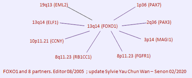
For alveolar rhabdomyosarcoma
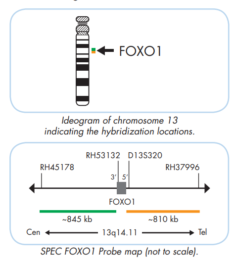
Orange Probe: telomere side, FOXO1 5' end
Green Probe: centromere side, FOXO1 3' end
The ZytoLight SPEC FOXO1 Dual Color Break Apart Probe is designed for the detection of specific translocations involving the chromosomal region 13q14.11 harboring the FOXO1 gene characteristic for alveolar rhabdomyosarcoma.
adapted from "Atlas of Genetics and Cytogenetics in Oncology and Haematology"
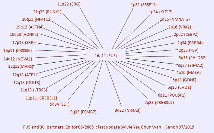
for myxoid liposarcoma.
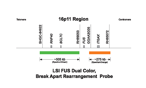
Orange Probe: centromere side, FUS, 3' end
Green Probe: telomere side, FUS, 5' end
Vysis LSI FUS Break Apart FISH Probe Kit is designed to detect translocations involving the chromosomal region 16p11.2 harboring the FUS gene. FUS gene rearrangements have been shown to be involved in both solid tumors and leukemias, e.g. in over 90% of myxoid liposarcoma.
adapted from "Atlas of Genetics and Cytogenetics in Oncology and Haematology"
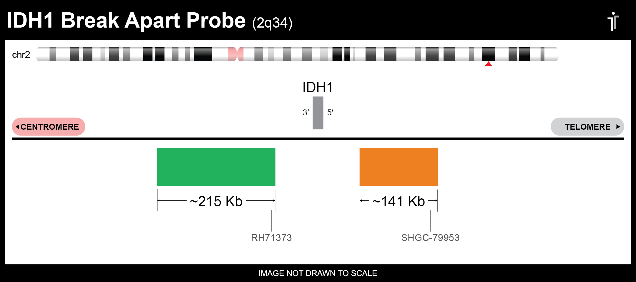
Orange Probe: telomere side, IDH1 5' end
Green Probe: cenromere side, IDH1 3' end
Empire Genomics’ IDH1 Break Apart FISH Probe is designed to flank the CDK6 gene and is typically used for detecting IDH1 rearrangements such as translocations.
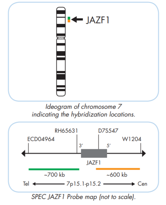
for low grade/high grade endometrial stromal sarcoma.
Orange Probe: centromere side, JAZF1 5' end
Green Probe: telomere side, JAZF1 3' end
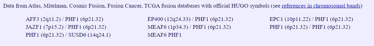
The ZytoLight ® SPEC JAZF1 Dual Color Break Apart Probe is designed to detect translocations involving the chromosomal region 7p15.1-p15.2 harboring the JAZF1 gene. In 25-30% of endometrial stromal sarcoma, espectially the low grade ones, the JAZF1 gene is disrupted by another translocation t(6;7) where the first zinc finger domain of JAZF1 is fused to both zinc finger domains of the PHF1 gene at 6p21.32.
adapted from "Atlas of Genetics and Cytogenetics in Oncology and Haematology"
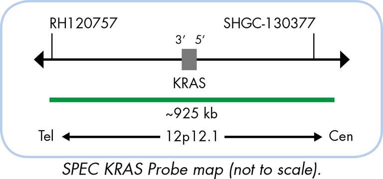
The ZytoLight ® SPEC KRAS/CEN 12 Dual Color Probe is designed for the detection of KRAS gene amplifications found e.g. in lung cancer. Mutations of KRAS are frequently found in epithelial malignancies and lead to activation of the downstream mitogen-activated protein kinase resulting in unchecked cellular proliferation and tumor progression.
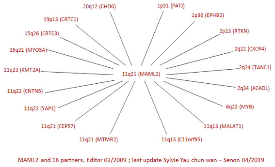
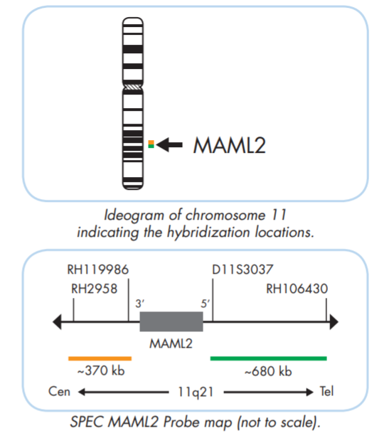
for mucoepidermoid carcinoma.
Orange Probe: centromere side, MAML2 3' end
Green Probe: telomere side, MAML2 5' end
The ZytoLight ® SPEC MAML2 Dual Color Break Apart Probe is designed to detect the translocation t(11;19)(q21;p13.1) specific for mucoepidermoid carcinomas.
adapted from "Atlas of Genetics and Cytogenetics in Oncology and Haematology"
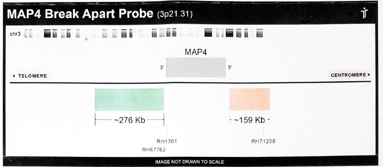
Orange Probe: centromere side, MAP4 5' end
Green Probe: telomere side, MAP4 3' end
For the detection of MAP4 re-arrangement (Chr.3p21.31). The protein encoded by this gene is a major non-neuronal microtubule-associated protein. This protein contains a domain similar to the microtubule-binding domains of neuronal microtubule-associated protein (MAP2) and microtubule-associated protein tau (MAPT/TAU). This protein promotes microtubule assembly, and has been shown to counteract destabilization of interphase microtubule catastrophe promotion. Cyclin B was found to interact with this protein, which targets cell division cycle 2 (CDC2) kinase to microtubules. The phosphorylation of this protein affects microtubule properties and cell cycle progression. Multiple transcript variants encoding different isoforms have been found for this gene.
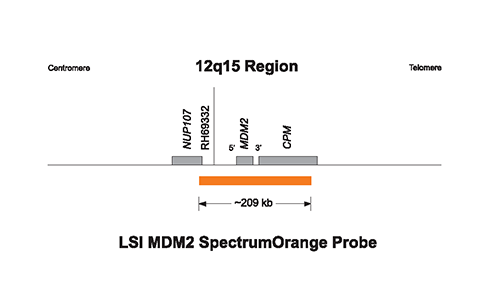
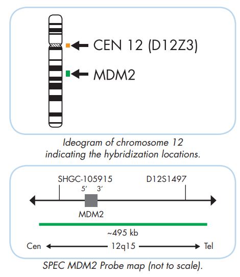
for well-differentiated liposarcoma and many others.
The Vysis MDM2/CEP 12 FISH Probe Kit and the ZytoLight ® SPEC MDM2/CEN 12 Dual Color Probe are designed for the detection of MDM2 gene amplifications. Well-differentiated liposarcomas (WDLPS) are characterized by the amplification of 12q-derived chromosomal material, harboring the MDM2 oncogene while lipomas show balanced translocations involving 12q13-15.
Please note that Vysis probe and Zytolight probe are of different colors:
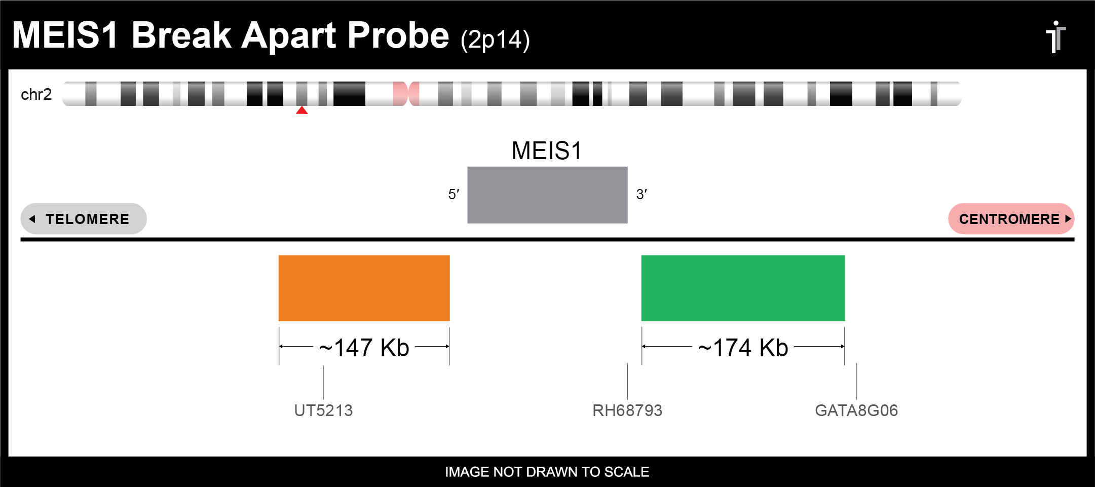
Orange Probe: telomere side, MEIS1 5' end
Green Probe: centromere side, MEIS1 3' end
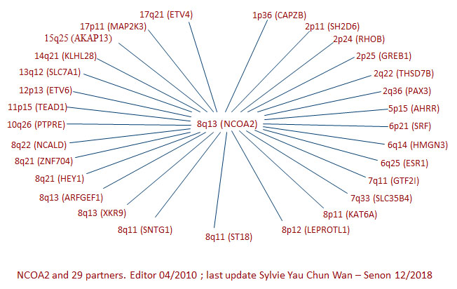
Empire Genomics’ MEIS1 Break Apart FISH Probe is designed to flank the MEIS1 gene and is typically used for detecting MEIS1 rearrangements such as translocations. Sarcomas with MEIS1-NCOA2 fusions have been reported in 2 cases each of primitive renal sarcomas and intraosseous pelvic rhabdomyosarcomas. Recurrent MEIS1-NCOA2/1 fusions in a subset of low-grade spindle cell sarcomas frequently involve the genitourinary and gynecologic tracts. The consistent morphology was that of monomorphic spindle to ovoid cells in a storiform, whorling, or solid pattern. Alternating cellularity, myxoid stroma, and microcystic changes were seen.
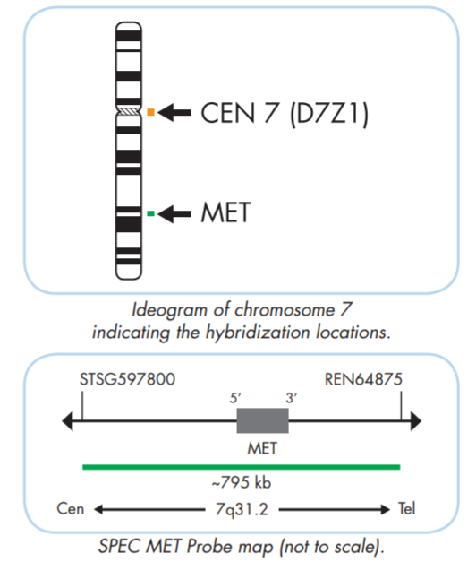
The ZytoLight ® SPEC MET/CEN 7 Dual Color Probe is designed for the detection of MET gene amplifications found in a variety of human tumors including lung, breast, colorectal, prostate and gastric carcinomas as well as in gliomas,melanomas and some sarcomas. Several inhibitors of the HGF/MET signaling pathway are being studied and developed as potent therapies to inhibit tumor growth.
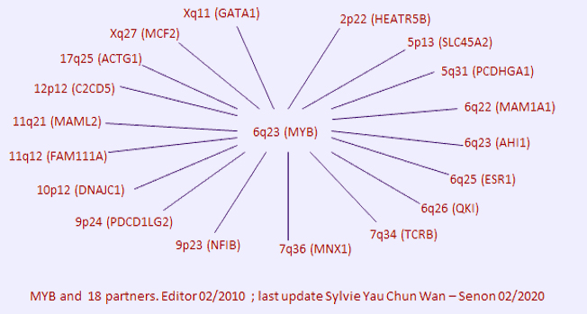
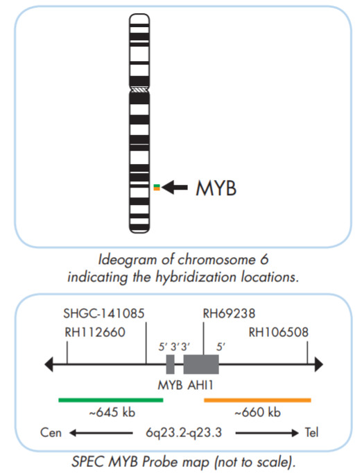
for adenoid cystic carcinoma.
Orange Probe: telomere side, MYB 3' end
Green Probe: centromere side, MYB 5' end
The ZytoLight ® SPEC MYB Dual Color Break Apart Probe is designed to detect translocations involving the chromosomal region 6q23.3 harboring the MYB gene. The MYB gene is expressed predominantly in immature progenitor cells of all hematopoietic lineages and is highly expressed in most leukemias and in some solid tumors. Translocations affecting MYB have been detected in T-cell acute lymphoblastic leukemia and adenoid cystic carcinoma.
adapted from "Atlas of Genetics and Cytogenetics in Oncology and Haematology"
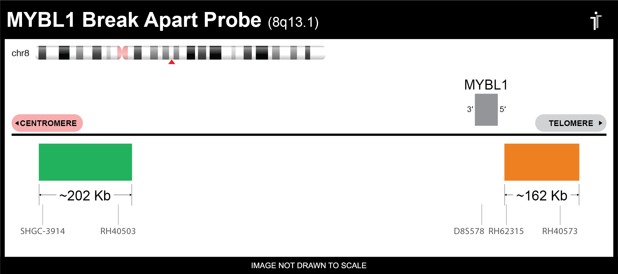
for adenoid cystic carcinoma.
Orange Probe: telomere side, MYBL1 5' end
Green Probe: centromere side, MYBL1 3' end
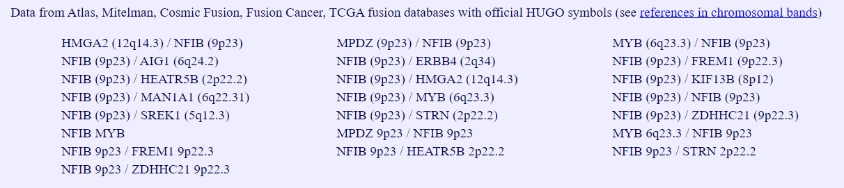
For adenoid cystic carcinoma, also consider MYB and NFIB break apart FISH tests. MYBL1 is one of those uncommon partners of NFIB.
Chromosome: CHR8: 67474409-67525480
Locus: 8q13.1
adapted from "Atlas of Genetics and Cytogenetics in Oncology and Haematology"
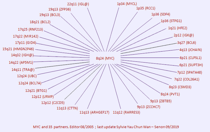
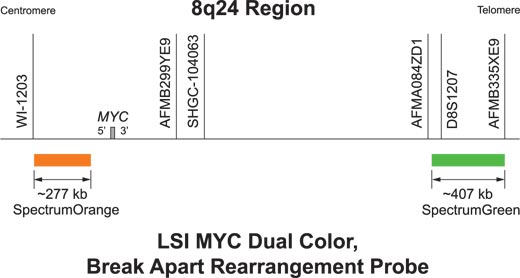
for double-hit, triple-hit diffuse large B-cell lymphoma.
Orange Probe: centromere side, MYC 5' end
Green Probe: telomere side, MYC 3' end
Vysis LSI MYC Break Apart Rearrangement Probe Kit is designed to detect translocations involving the chromosomal region 8q24.21 harboring the MYC gene. Translocations involving the MYC gene are considered to be cytogenetic hallmarks for Burkitt Lymphoma but are also found in other types of lymphomas.
In high-grade B-cell lymphomas, the presence of MYC aberrations identifies a patient subset with a very poor prognosis, particularly when there is concomitant rearrangement of BCL2 or BCL6, a condition referred to as "double hit” DLBCL. In rare cases translocation involves MYC, BCL2 and BCL6, so called “triple hit”. MYC translocations confer a worse prognosis in patients treated with cyclophosphamide, doxorubicin, vincristine, and prednisone (CHOP), both in combination with and without rituximab.
adapted from "Atlas of Genetics and Cytogenetics in Oncology and Haematology"
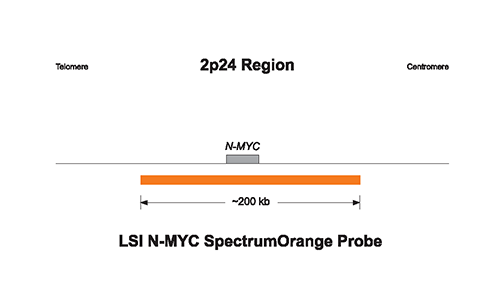
for neuroblastoma.
Orange Probe: MYCN, 2p24, ~200 kb
Vysis LSI N-MYC SpectrumOrange Probe is designed for the detection of MYCN amplification which represents the most powerful unfavorable prognostic factor for neuroblastoma. Less frequently amplifications are found in retinoblastoma, small cell lung cancer, astrocytoma and other tumors derived from the neuroectoderm. Amplification of the MYCN gene is found in about 25% of primary neuroblastomas and is strongly associated with rapid tumor progression, advanced stages of the disease, and poor prognosis.
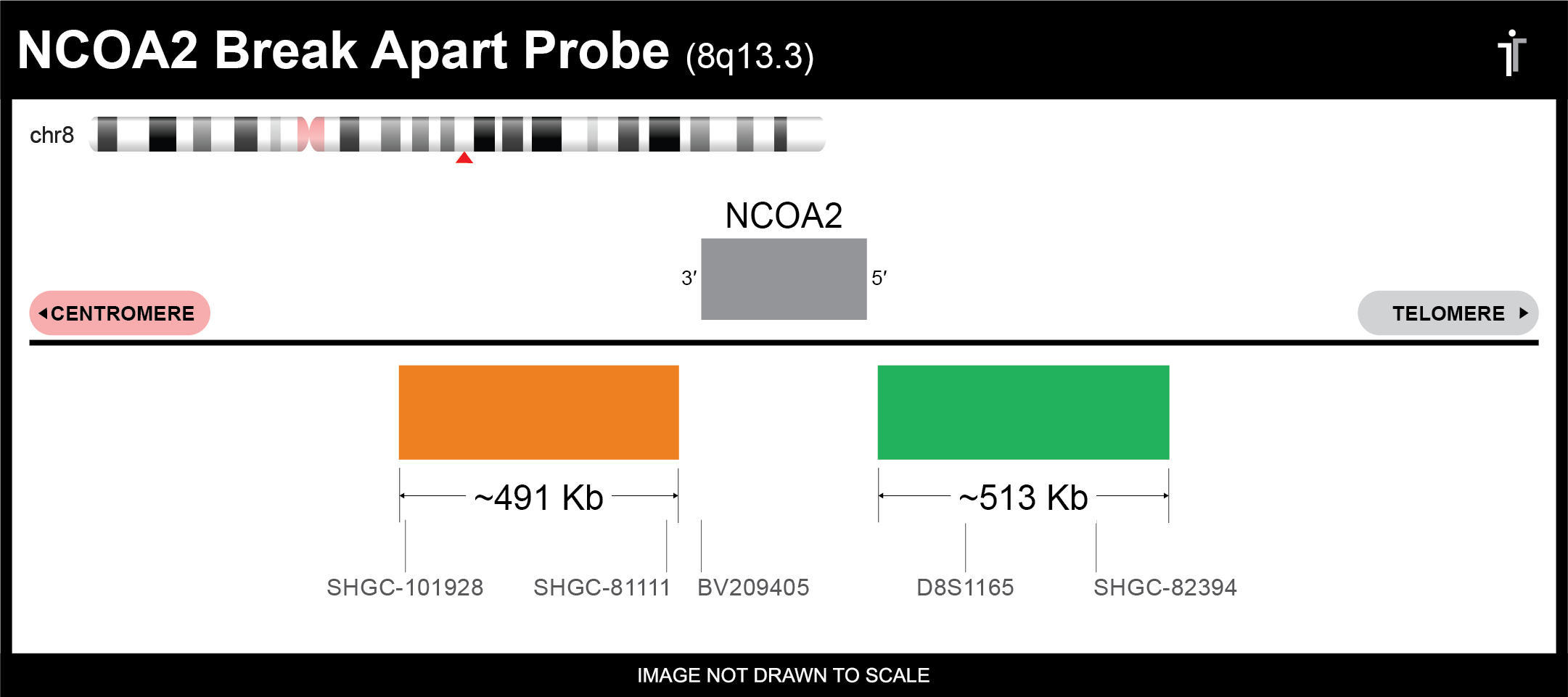
Orange Probe: centromere side, NCOA2 3' end
Green Probe: telomere side, NCOA2 5' end
adapted from "Atlas of Genetics and Cytogenetics in Oncology and Haematology"
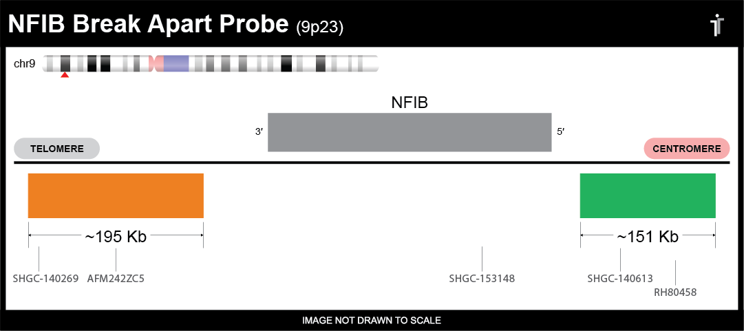
for adenoid cystic carcinoma.
Orange Probe: telomere side, NFIB 3' end
Green Probe: centromere side, NFIB 5' end
The t(6;9)(q22-23;p23-24) translocation in adenoid cystic carcinomas (ACC) of the breast and H&N consistently results in fusions encoding chimeric transcripts predominantly consisting of MYB exon 14 linked to the last coding exon(s) of NFIB. The minimal common part of MYB deleted as the result of fusion was exon 15 including the 3'-UTR, which contains several highly conserved target sites for miR-15a/16 and miR-150 microRNAs. PNAS 2009 106(44):18740-4.
adapted from "Atlas of Genetics and Cytogenetics in Oncology and Haematology"
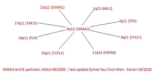
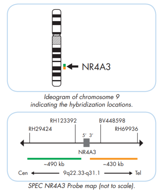
for extraskeletal myxoid chondrosarcoma.
Orange Probe: telomere side, NR4A3 3' end
Green Probe: centromere side, NR4A3 5' end
The ZytoLight ® SPEC NR4A3 Dual Color Break Apart Probe is designed to detect translocations involving the chromosomal region 9q22.33-q31.1. The most frequent reciprocal translocation is t(9;22)(q22.3-q31;q12.2) found in about 70% of extraskeletal myxoid chondrosarcoma generating a EWSR1-NR4A3 fusion gene in which the 3’-terminal part of EWSR1 is replaced by the entire NR4A3 gene. Extraskeletal myxoid chondrosarcoma is histologically characterized by a mixture of cellular and myxoid stromal components, making it difficult to distinguish it from other benign or malignant mesenchymal tumors, especially those with myxoid matrix.
adapted from "Atlas of Genetics and Cytogenetics in Oncology and Haematology"
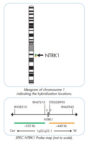
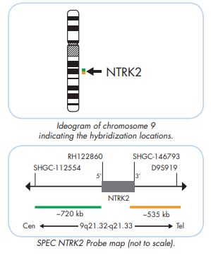
for pan cancer biomarkers.
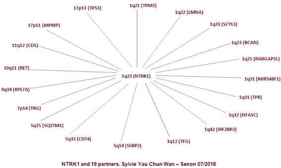
To detect translocations involving the chromosomal regions 1q23.1 harboring the NTRK1 gene, 9q21.33 harboring the NTRK2 gene, and 15q25.3 harboring the NTRK3 gene, respectively. Tumors presenting NTRK1, NTRK2 and NTRK3 gene fusions can be sensitive to targeted therapies with tyrosine kinase inhibitors. The treatment of patients with NTRK1, 2, or 3 fusion-positive cancers with an NTRK inhibitor, such as the FDA-approved drugs larotrectinib or entrectinib, is associated with high response rates, regardless of NTRK gene, fusion partner, and tumor type.
Mixed TRK IHC might be used for preliminary screening.
NTRK1 rearrangements were shown to be involved in many different tumor types in which more than 40 different fusion partners have been described, resulting in an activated chimeric NTRK1 gene.
adapted from "Atlas of Genetics and Cytogenetics in Oncology and Haematology"
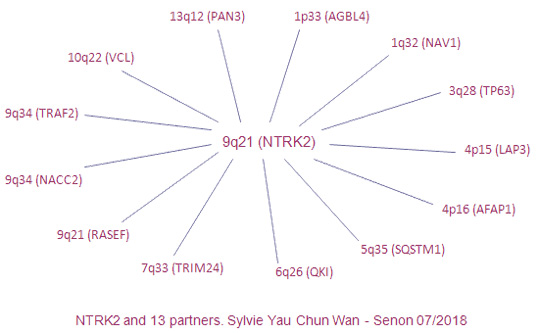
NTRK2 is a tyrosine kinase and plays a key role in central and peripheral nervous system development as well as in cell survival. Translocations affecting the NTRK2 gene have been reported in several cancer types, including glioblastomas, pilocytic astrocytomes, H&N squamous cell carcinoma, and lung adenocarcinoma.
adapted from "Atlas of Genetics and Cytogenetics in Oncology and Haematology"
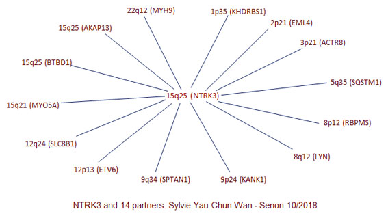
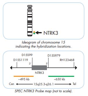
Translocations affecting the NTRK3 gene have been reported in several cancer types, including glioblastomas, Philadelphia chromosome-like acute lymphoblastic leukemia, congenital fibrosarcomas, cellular mesoblastic nephromas, acute myeloid leukemia, radiation-associated thyroid cancer, secretory breast carcinoma, and mammary analog secretory carcinoma of the salivary gland.
adapted from "Atlas of Genetics and Cytogenetics in Oncology and Haematology"
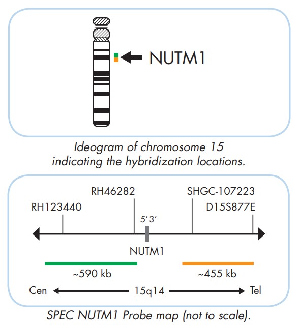
Orange Probe: telomere side, NUTM1 3' end
Green Probe: centromere side, NUTM1 5' end
The ZytoLight ® SPEC NUTM1 Dual Color Break Apart Probe is designed to detect translocations involving the chromosomal region 15q14 harboring the NUTM1 gene. NUT midline carcinoma (NMC) is a rare and aggressive form of squamous cell carcinoma that arises mainly in the head, neck, or mediastinum. NMCs may be indistinguishable from more common squamous cell carcinomas and are thus an underdiagnosed entity. Therefore, the diagnosis of NMC depends on the confirmation of NUTM1 rearrangement.
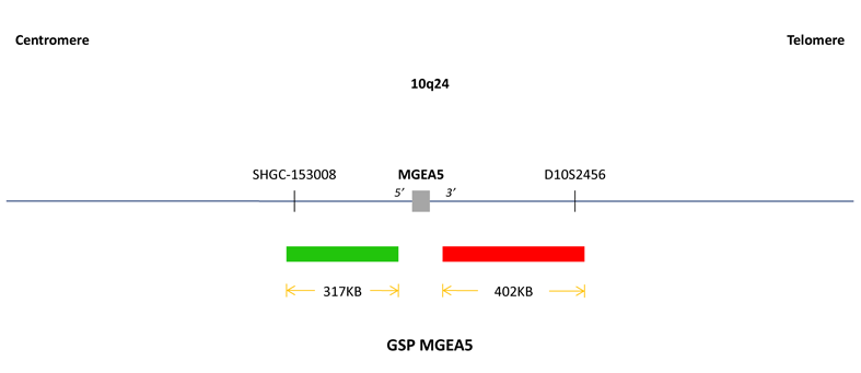
Orange Probe: telomere side, OGA 3' end
Green Probe: centromere side, OGA 5' end
TGFBR3/MGEA5 t(1;10)(p22;q24) might be present in Hemosiderotic Fibrolipomatous Tumour (HFLT), Pleomorphic Hyalinizing Angiectatic Tumor (PHAT) and Myxoinflammatory Fibroblastic Sarcoma (MIFS), etc.
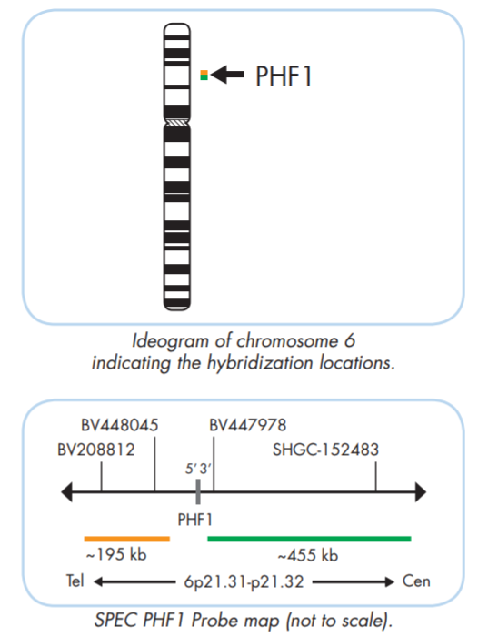
for low grade endometrial stroma tumor.
Orange Probe: telomere side, PHF1 5' end
Green Probe: centromere side, PHF1 3' end
The ZytoLight ® SPEC PHF1 Dual Color Break Apart Probe is designed for the detection of translocations involving the chromosomal region 6p21.32 harboring the PHF1 gene. Endometrial stromal tumors are the second most common pure mesenchymal tumors of the uterus.
adapted from "Atlas of Genetics and Cytogenetics in Oncology and Haematology"
Several rearrangements involving the genes BCOR, JAZF1, PHF1, or YWHAE have been identified in endometrial stromal tumors.
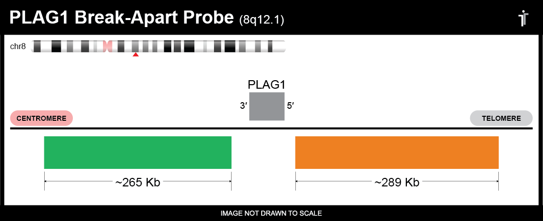
Orange Probe: telomere side, PLAG1 5' end
Green Probe: centromere side, PLAG1 3' end
Pleomorphic adenoma gene 1.
For pleomorphic adenoma, chondroid syringoma, myoepithelial tumors of skin and soft tissue, lipoblastoma, pediatric fibromyxoid soft tissue tumor, PLAG mesenchymal tumours or “plagomas”, certain uterine leiomyosarcoma, etc.
adapted from "Atlas of Genetics and Cytogenetics in Oncology and Haematology"
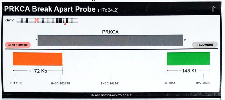
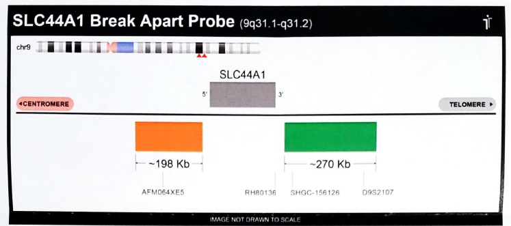
Orange Probe: centromere side, PRKCA 5' end
Green Probe: telomere side, PRKCA 3' end
Mixed neuronal-glial tumors are rare and challenging to subclassify. One recognized variant, papillary glioneuronal tumor (PGNT), is characterized by prominent pseudopapillary structures and glioneuronal elements. The t(9;17)(q31;q24) with the resultant novel fusion oncogene SLC44A1-PRKCA is the defining molecular feature of PGNT that may be responsible for its pathogenesis. Bridge et al Brain Pathol 2013 PMID: 22725730.
SLC44A1 break apart FISH and RT-PCR for the fusion transcript SLC44A1-PRKCA are also available in the lab.
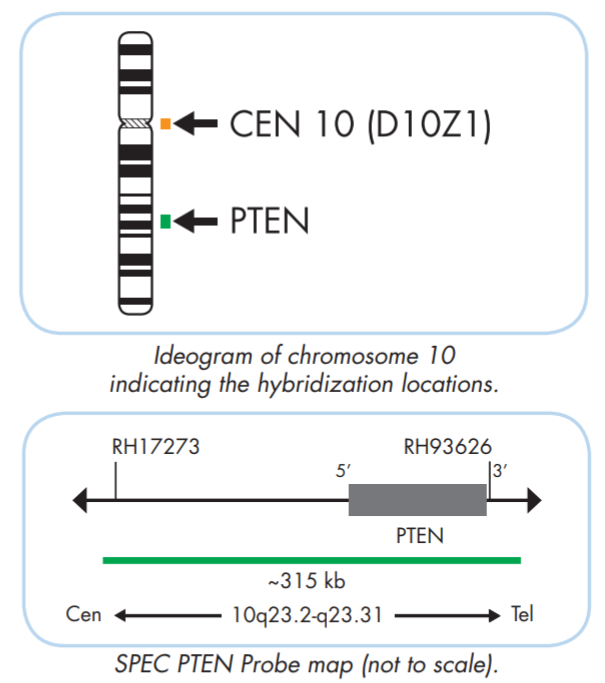
The ZytoLight ® SPEC PTEN/CEN 10 Dual Color Probe or is designed for the detection of PTEN deletions frequently observed in many tumor types, including renal, melanoma, endometrial, breast, prostate, lung, bladder, and thyroid cancer but also in hematological neoplasms. Deletions affecting the long arm of chromosome 10 have been detected in 30 to 50% of early and advanced stage sporadic melanomas and about 40 to 70% of prostate cancers. This probe can also be used for prognostic evaluation and in the diagnostic algorithm of glioma.
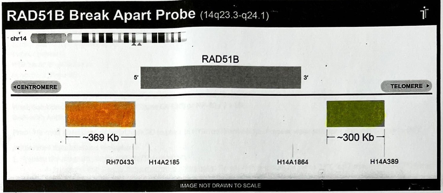
Orange Probe: centromere side, RAD51B 5' end
Green Probe: telomere side, RAD51B 3' end
might be uterine leiomyoma and some other sarcomas.
adapted from "Atlas of Genetics and Cytogenetics in Oncology and Haematology"
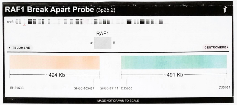
Orange Probe: telomere side, RAF1 3' end
Green Probe: centromere side, RAF1 5' end
A subset of spindle cell tumours have been recently identified to harbor recurrent fusion genes, involving NTRK1/2/3, BRAF, RAF1, and RET. The precise classification of these fusion-positive tumours relies essentially on genomic profiling.
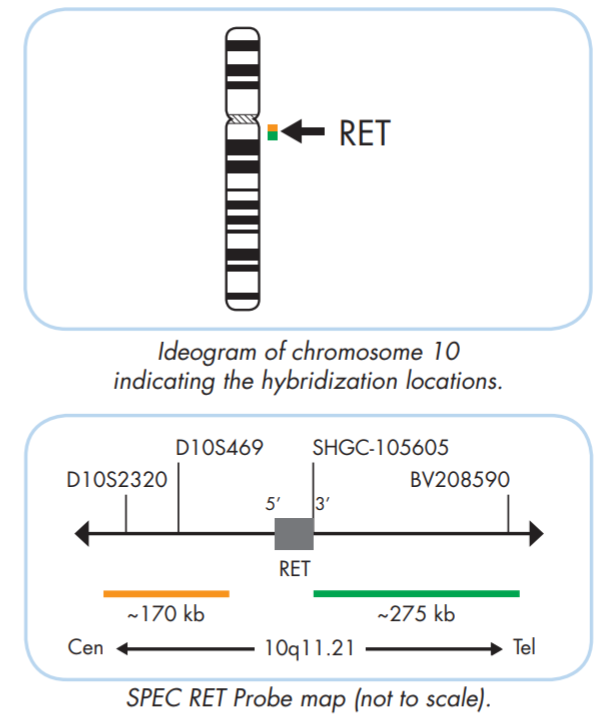
potential pan cancer biomarker.
Orange Probe: centromere side, RET 5' end
Green Probe: telomere side, RET 3' end
The ZytoLight ® SPEC RET Dual Color Break Apart Probe is designed to detect translocations involving the chromosomal region 10q11.21 harboring the RET gene observed e.g. in lung adenocarcinoma.
Since in vitro studies showed transforming activity of KIF5B-RET fusions which could be suppressed by a tyrosine kinase inhibitor, it was assumed that the chimeric oncogene might be a promising molecular target for the treatment of lung cancer. The cut off is set as low as 15% tumor nuclei.
Salivary glands: mammary analog secretory carcinoma
ERC1
12q13.33 (991208 )
t(10;12)(q11;q13) ERC1/RET
Breast cancer
Lung: non-small cell lung carcinoma
Thyroid: papillary thyroid carcinoma
ANKS1B
12q23.1 (98743974)
t(10;12)(q11;q23) ANKS1B/RET
Lung: non-small cell lung carcinoma
CLIP1
12q24.31 (122271434)
t(10;12)(q11;q24) CLIP1/RET
Lung: non-small cell lung carcinoma
TSSK4
14
14q12 (24205720)
t(10;14)(q11;q12) TSSK4/RET
Lung: non-small cell lung carcinoma
KTN1
14q22.3 (55580207)
t(10;14)(q11;q22) KTN1/RET
Thyroid: papillary thyroid carcinoma
CCDC88C
14q32.11 (91271323)
t(10;14)(q11;q32) CCDC88C/RET
Lung: non-small cell lung carcinoma
GOLGA5
14q32.12 (92794231)
t(10;14)(q11;q32) GOLGA5/RET
Skin: melanomas/Spitz tumors
Thyroid: papillary thyroid carcinoma
MYO5C
15
15q21.2 (52192318)
t(10;15)(q11;q21) MYO5C/RET
Lung: non-small cell lung carcinoma
AKAP13
15q25.3 (85380616)
t(10;15)(q11;q25) AKAP13/RET
Thyroid: papillary thyroid carcinoma
MYH10
17
17p13.1 (8474205)
t(10;17)(q11;p13) MYH10/RET
Soft tissues: Infantile myofibromatosis
Soft tissues: spindle mesenchymal neoplasm
MYH13
17p13.1 (10300866)
t(10;17)(q11;p13) MYH13/RET
Thyroid: papillary thyroid carcinoma
MPRIP
17p11.2 (17042760)
t(10;17)(q11;p11) MPRIP/RET
Lung: non-small cell lung carcinoma
PRKAR1A
17q24.2 (68512379)
t(10;17)(q11;q24) PRKAR1A/RET
Lung: non-small cell lung carcinoma
Neuro-endocrine tumor
RELCH (KIAA1468)
18
18q21.33 (62187291)
t(10;18)(q11;q21) KIAA1468/RET
Lung: adenocarcinoma
Lung: non-small cell lung carcinoma
Thyroid: papillary thyroid carcinoma
LSM14A
19
19q13.11 (34172447)
t(10;19)(q11;q13) LSM14A/RET
Lung: adenocarcinoma
RRBP1
20
20p12.1 (17613678)
t(10;20)(q11;p12) RRBP1/RET
Colorectal cancer
BCR
22
22q11.23 (23180365)
t(10;22)(q11;q11) BCR/RET
Chronic myeloproliferative neoplasm
SPECC1L
22q11.23 (24270817)
t(10;22)(q11;q11) SPECC1L/RET
Thyroid: papillary thyroid carcinoma
TIMP3
22q12.3 (32800816)
t(10;22)(q11 ;q12) TIMP3/RET
Soft tissues: Inflammatory myofibroblastic tumor
adapted from "Atlas of Genetics and Cytogenetics in Oncology and Haematology"
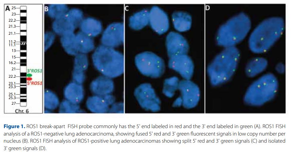
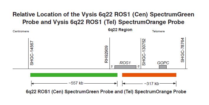
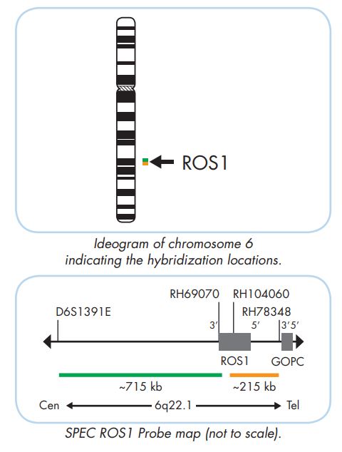
for non-small cell lung carcinoma (CAP accredited test).
Orange Probe: telomere side, ROS1 5' end
Green Probe: centromere side, ROS1 3' end
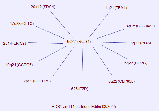
The Vysis ROS1 Break Apart FISH Probe Kit or the ZytoLight ® SPEC ROS1 Dual Color Break Apart Probe is designed to detect ROS1 gene rearrangements at 6q22.1 involving the receptor tyrosine kinase (ROS1) gene. Rearrangement of the ROS1 gene occurs in:
- 0.2%-2.4% of Colorectal Cancers (CRC),
- 1%-2% of Non-Small Cell Lung Cancer (NSCLC) leading to its oncogenic activation
- Gastric Adenocarcinoma
- Various types of melanoma, inflammatory myofibroblastic tumor, and angiosarcoma
- Epithelioid hemangioendothelioma.
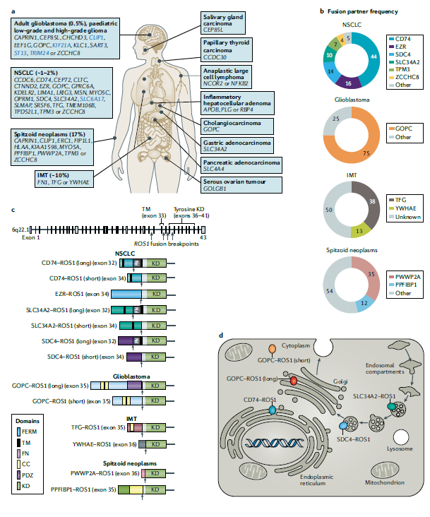
a) ROS1 fusions have been identified in several cancer types that occur in adults and/or children (shown on a body map). Upstream gene partners of ROS1 fusions found in specific cancer types are listed. Among these, CLIP1, KIF21A, ST13, TRIM24 and SLC6A17 (blue font) were identified within the cBioPortal database but have not been reported in peer-reviewed publications. In terms of the absolute number of patients affected, ROS1 fusions are most often found in non-small-cell lung cancers (NSCLCs) given the substantial global burden of this disease relative to that of other malignancies. According to data from the cBioPortal, 78% of patients found to have a ROS1 fusion in their cancer had lung adenocarcinoma, with other cancers accounting for the remaining 22%. The prevalence of ROS1 fusions is higher among certain rare cancers such as Spitzoid neoplasms and inflammatory myofibroblastic tumours (IMTs), which affect a smaller total number of patients as compared with NSCLC. The reported prevalence of ROS1 fusions is indicated for certain cancers in parentheses in the Figure. b) ROS1 fusion partner frequencies in ROS1-rearranged NSCLCs, adult glioblastomas, IMTs and Spitzoid neoplasms are shown in circular plots (percentages shown). Large-cohort studies are lacking for other cancer types, and frequency data are thus unavailable. See Supplementary Table 1 for data on median or aggregate frequencies for each cancer type. c) The locations of four major intronic breakpoints (within introns 31, 33, 34 or 35) that generate ROS1 fusions are indicated by arrows in the upper panel. The domain organization of recurrent ROS1 fusions in NSCLC, glioblastoma, IMT and Spitz tumours is shown in the lower panel. An intact ROS1 tyrosine kinase domain (KD) and the C-terminal domain (corresponding to exons 36–43) are included in all fusions. However, ROS1 fusions can also include a portion of the last fibronectin type III (FN) motif repeat (exon 32), the transmembrane (TM) domain (exon 33) and/or a portion of the juxtamembrane domain (exons 34 and 35) of ROS1; retention of portions of these domains does not seem to affect oncogenicity. The majority of NSCLC-associated ROS1 fusion partners lack dimerization motifs, suggesting that canonical dimerization might not be required for oncogenic activation. Distinct from the native receptor, ROS1 fusions might be activated simply by conformational changes induced by the removal of most of the extracellular domain and redirection of this activity to novel subcellular locations. d) The subcellular localizations of select ROS1 fusion proteins are depicted. CC, coiled-coil domain; FERM, band 4.1 homology and ezrin–radixin–moesin domain; PDZ, PSD95, Dlg1 and ZO-1 domain. (Drilon et al, 2021, Nature Reviews Clinical Oncology (18), pp.35–55.)
See also "IASLC Atlas of ALK adn ROS1 testing in lung cancer 2e".
adapted from "Atlas of Genetics and Cytogenetics in Oncology and Haematology"
Vysis Probe
ZytoLight Probe
Orange Probe: centromere side, SLC44A1 5' end
Green Probe: telomere side, SLC44A1 3' end
Mixed neuronal-glial tumors are rare and challenging to subclassify. One recognized variant, papillary glioneuronal tumor (PGNT), is characterized by prominent pseudopapillary structures and glioneuronal elements. The t(9;17)(q31;q24) with the resultant novel fusion oncogene SLC44A1-PRKCA is the defining molecular feature of PGNT that may be responsible for its pathogenesis. Bridge et al Brain Pathol 2013 PMID: 22725730.
PRKCA break apart FISH and RT-PCR for the fusion transcript SLC44A1-PRKCA are also available in the lab.
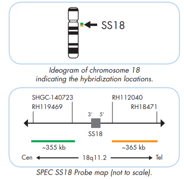
for synovial sarcoma.
Orange Probe: telomere side, SS18 5' end
Green Probe: centromere side, SS18 3' end
The ZytoLight ® SPEC SS18 Dual Color Break Apart Probe is designed to detect translocations involving the chromosomal region 18q11.2 harboring the SS18 gene. Translocations involving the region 18q11.2 are found in over 90% of synovial sarcoma.
adapted from "Atlas of Genetics and Cytogenetics in Oncology and Haematology"
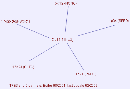
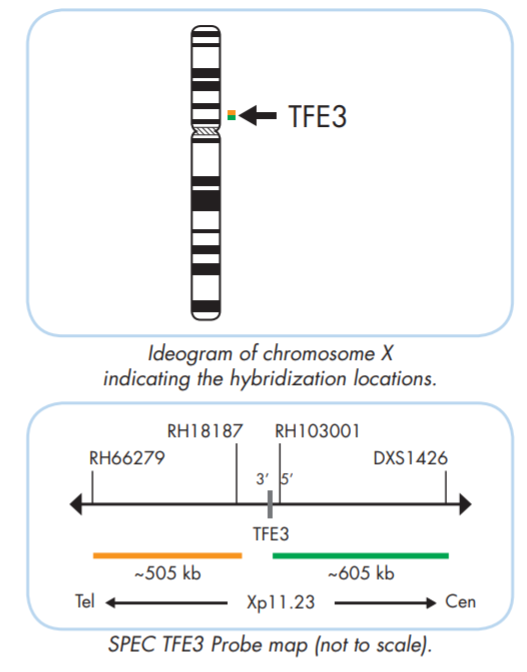
for papillary RCC and alveolar soft part sarcoma.
Orange Probe: centromere side, TFE3 3' end
Green Probe: telomere side, TFE3 5' end
The ZytoLight ® SPEC TFE3 Dual Color Break Apart Probe is designed to detect translocations involving the chromosomal region Xp11.23 harboring the TFE3 gene. Translocations involving the chromosomal region Xp11.2 are frequently detected in some forms of renal cell carcinomas (e.g. papillary RCC), and in alveolar soft part sarcoma.
adapted from "Atlas of Genetics and Cytogenetics in Oncology and Haematology"
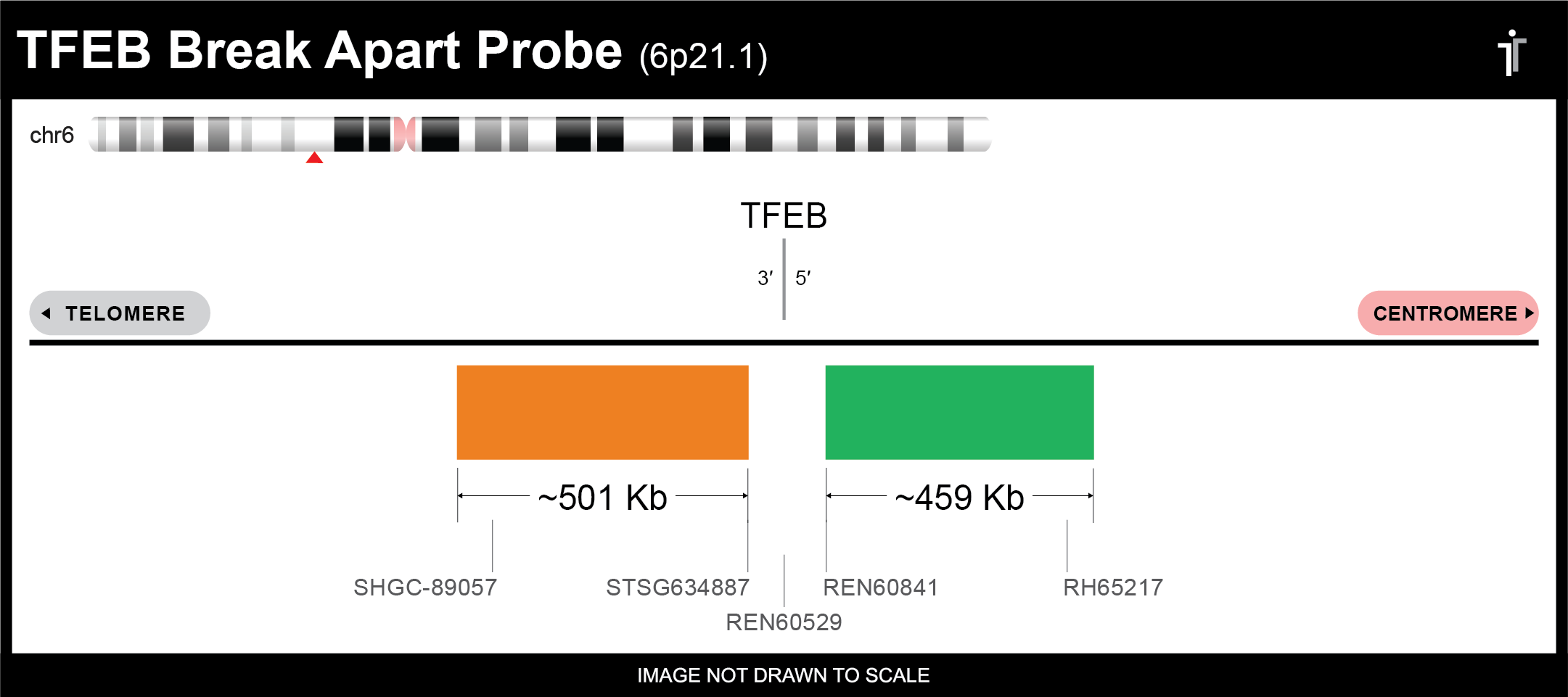
Orange Probe: telomere side, TFEB 3' end
Green Probe: centromere side, TFEB 5' end
For RCC typing.
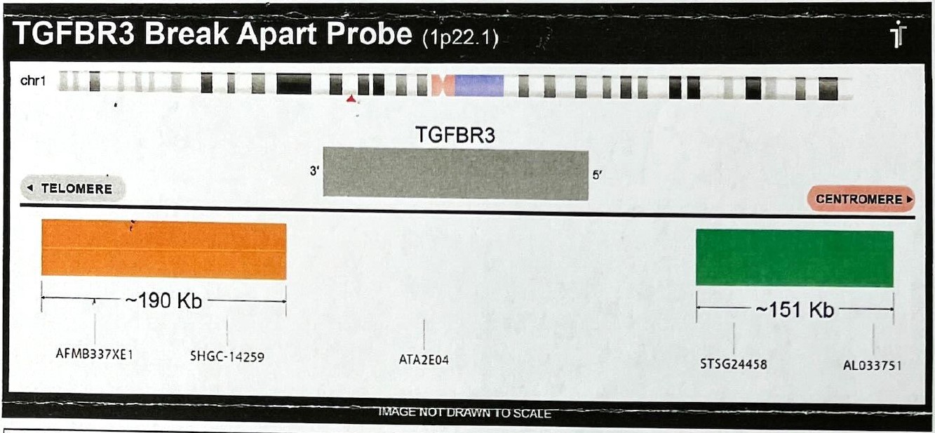
Orange Probe: teloomere side, TGFBR3 5' end
Green Probe : centromere side, TGFBR3 3' end
might be many sarcomas, e.g. Myxoinflammatory Fibroblastic Sarcoma and Hemosiderotic Fibrolipomatous Tumor.
adapted from "Atlas of Genetics and Cytogenetics in Oncology and Haematology"
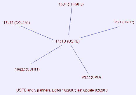
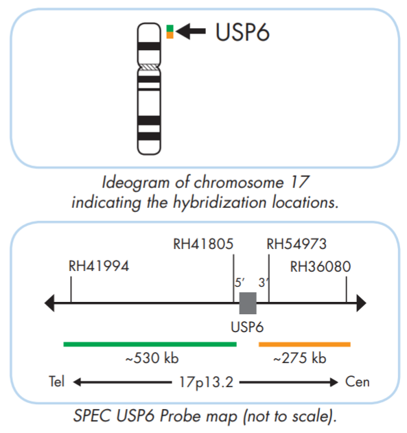
for nodular fasciitis and primary ABC, and the group of USP6-rearranged lesions (giant cell lesion of the small bones, myositis ossificans, fibro-osseous pseudotumor of digits and fibroma of tendon shealth)
Orange Probe: centromere side, USP6 3' end
Green Probe: telomere side, USP6 5' end
The ZytoLight ® SPEC USP6 Dual Color Break Apart Probe is designed to detect translocations involving the USP6 gene, at chromosome location, 17p13.2. Translocations affecting USP6 have been found in primary aneurysmal bone cysts (ABC) and nodular fasciitis (NF).
adapted from "Atlas of Genetics and Cytogenetics in Oncology and Haematology"
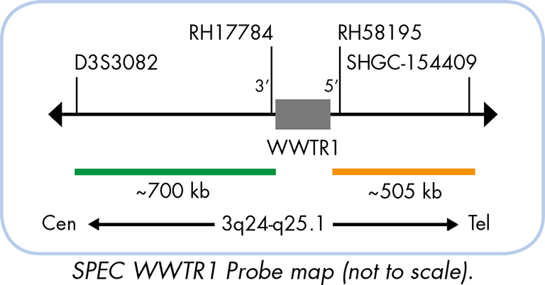
for epithelioid hemangioendothelioma.
Orange Probe: telomere side, WWTR1 5' end
Green Probe: centromere side, WWTR1 3' end
The ZytoLight ® SPEC WWTR1 Dual Color Break Apart Probe is designed for the detection of translocations involving the chromosomal region 3q25.1 harboring the WWTR1 gene. The recurrent translocation t(1;3)(p36.3;q25.1) was identified in approximately 90% of epithelioid hemangioendothelioma (EHE) cases, but not in other vascular tumors. Another known fusion is YAP1-TFE3, which can co-exist with WWTR1-CAMTA1 translocation.
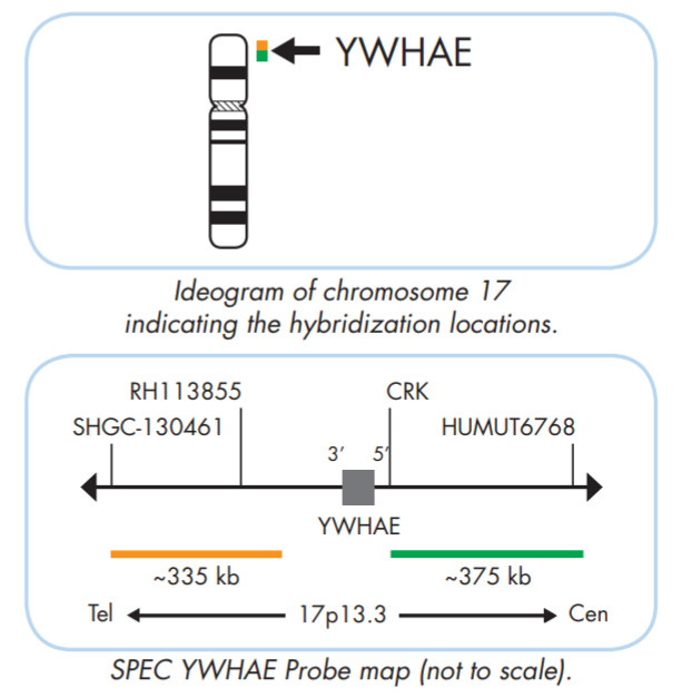
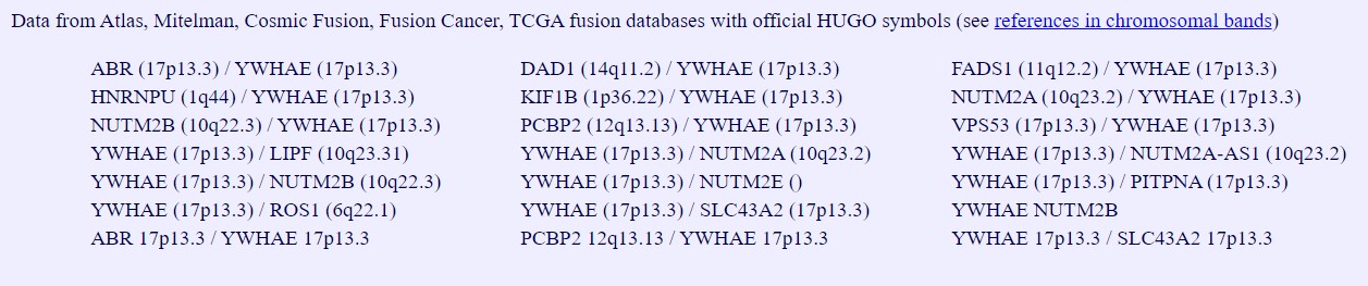
for high-grade endometrial stromal sarcoma.
Orange Probe: telomere side, YWHAE 3' end
Green Probe: centromere side, YWHAE 5' end
The ZytoLight ® SPEC YWHAE Dual Color Break Apart Probe is designed to detect rearrangements involving the chromosomal region 17p13.3 harboring the YWHAE gene. Altered expression of 14-3-3 family proteins is associated with development and progression of cancer. The fusion between YWHAE and one of the FAM22 family members (FAM22A or FAM22B) caused by a t(10;17)(q22;p13) has been identified in high-grade endometrial stromal sarcoma (ESS) as well as in clear cell sarcoma of the kidney (CCSK).
Official Full Name: YWHAE tyrosine 3-monooxygenase/tryptophan 5-monooxygenase activation protein epsilon
Locus: 17p13.3
adapted from "Atlas of Genetics and Cytogenetics in Oncology and Haematology"


 adapted from "Atlas of Genetics and Cytogenetics in Oncology and Haematology"
-Dual-Color-Break-Apart-Rearrangement-Probe_ProbeMap.jpg)
 adapted from "Atlas of Genetics and Cytogenetics in Oncology and Haematology"
 adapted from "Atlas of Genetics and Cytogenetics in Oncology and Haematology"
 adapted from "Atlas of Genetics and Cytogenetics in Oncology and Haematology"
 adapted from "Atlas of Genetics and Cytogenetics in Oncology and Haematology"
 adapted from "Atlas of Genetics and Cytogenetics in Oncology and Haematology"
 adapted from "Atlas of Genetics and Cytogenetics in Oncology and Haematology"
 adapted from "Atlas of Genetics and Cytogenetics in Oncology and Haematology"
 adapted from "Atlas of Genetics and Cytogenetics in Oncology and Haematology"
 adapted from "Atlas of Genetics and Cytogenetics in Oncology and Haematology"
 adapted from "Atlas of Genetics and Cytogenetics in Oncology and Haematology"
 adapted from "Atlas of Genetics and Cytogenetics in Oncology and Haematology"
 adapted from "Atlas of Genetics and Cytogenetics in Oncology and Haematology"
 adapted from "Atlas of Genetics and Cytogenetics in Oncology and Haematology"
 adapted from "Atlas of Genetics and Cytogenetics in Oncology and Haematology"
 adapted from "Atlas of Genetics and Cytogenetics in Oncology and Haematology"
 adapted from "Atlas of Genetics and Cytogenetics in Oncology and Haematology"
 adapted from "Atlas of Genetics and Cytogenetics in Oncology and Haematology"
 adapted from "Atlas of Genetics and Cytogenetics in Oncology and Haematology"
 adapted from "Atlas of Genetics and Cytogenetics in Oncology and Haematology"
 adapted from "Atlas of Genetics and Cytogenetics in Oncology and Haematology"
 adapted from "Atlas of Genetics and Cytogenetics in Oncology and Haematology"
 adapted from "Atlas of Genetics and Cytogenetics in Oncology and Haematology"
 adapted from "Atlas of Genetics and Cytogenetics in Oncology and Haematology"
 adapted from "Atlas of Genetics and Cytogenetics in Oncology and Haematology"
 adapted from "Atlas of Genetics and Cytogenetics in Oncology and Haematology"
 adapted from "Atlas of Genetics and Cytogenetics in Oncology and Haematology"
 adapted from "Atlas of Genetics and Cytogenetics in Oncology and Haematology"
 adapted from "Atlas of Genetics and Cytogenetics in Oncology and Haematology"